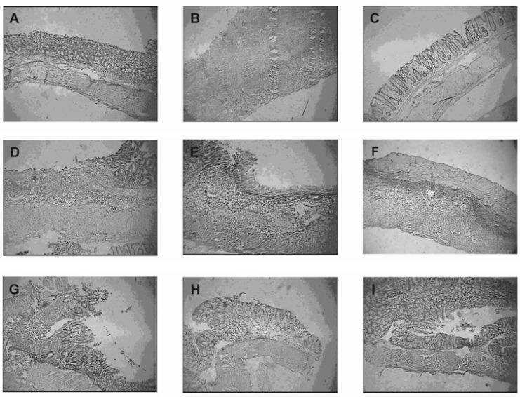
Figure 5.
Representative photographs of histological appearance of rat colonic mucosa. A= Normal control group, B= Colitis control group, after exposure to TNBS (100 mg/kg) the colon is markedly inflamed, the mucosal wall is thickened, and there is a transmural inflammatory cell infiltration. C= Budesonide pectin/Surelease coated pellet (300 μg/kg/day) group, D= Budesonide solution group (300 μg/kg/day), E=Budesonide uncoated pellet group (300 μg/kg/day), F= Placebo pellet group, G= Mesalazine enema (400 mg / kg/day, rectally) group, H= Budesonide enema (20 mcg/kg/day, rectally) group and I= Prednisolone (5 mg /kg /day, oral) group. Hematoxylin and eosin stain with original magnification 10×.

