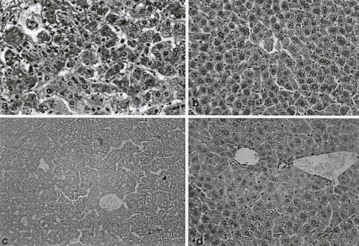
Figure 3.
Photomicrograph of lobules from control groups and fruit extract of Feijoa sellowiana at differences concentration–treated liver. Staining of neagative Control shows that cytoplasm was acidophilic and surrounded by a bright basophilic nucleus (a). MDMA (5 mg/kg) or positive control showed limited changes in lobules of liver and hepatocellular necrosis, with infiltration of mononuclear cells and accumulation of necrotic Kupffer cells with Pyknotic nuclei (b). Histo-pathological changes of aqueous (for comparison) and methanolic extract of Feijoa sellowiana fruit at a single dose of 100 mg/kg, respectively (c and d).

