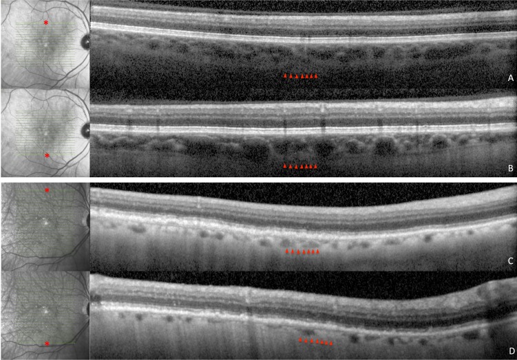
Figure 4.
Superior and inferior spectral domain optical coherence tomography images and corresponding infrared images of patients with AMD with and without RPD. Choroidal thickness in the non-RPD patient is 231 μm superiorly (A) and 242 μm inferiorly (B) and in the RPD patient is 107 μm superiorly (C) and 82 μm inferiorly (D). Point of measurement on macula is denoted by an asterisk in infrared images.

