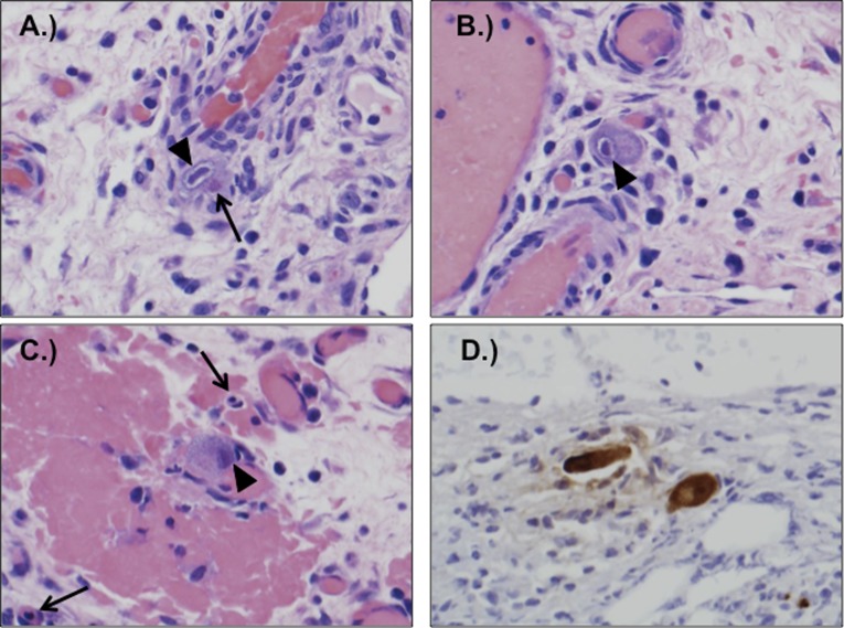
FIGURE 1.
A–C, Sections of surgically resected bowel showed enlarged endothelial cells with viral cytopathic changes, including eosinophilic cytoplasmic inclusions and basophilic nuclear inclusions. Arrows point to cytoplasmic (open arrows) and nuclear (arrowheads) inclusions. D, Immunoperoxidase staining with anti-CMV antibody was positive in affected cells.

