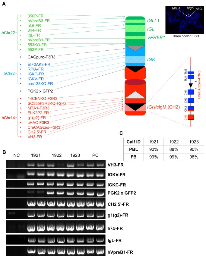
Figure 9. Establish DT40 clones carrying the engineered cKSL-HACΔ.
(A), Diagram of cKSL-HACΔ and the locations of PCR primers used to examine cKSL-HACΔ structural integrity. (B), A representative gel image of PCR products for some of the junction points in the cKSL-HACΔ from genomic DNA isolated from PBL of Tc claves 1921, 1922 and 1923. (C) cKSL-HACΔ retention rates in Tc calves 1921, 1922 and 1923 analyzed by FISH. PBL: peripheral blood lymphocytes; FB: fibroblasts.

