
Fig. 4. REGγ mediates degradation of SirT1.
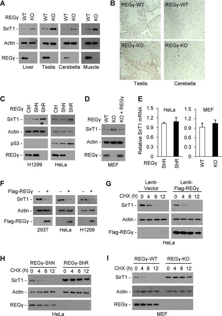
(A–E) REGγ deficiency causes accumulation of endogenous SirT1. (A–B) SirT1 protein levels in liver, testis, cerebellum and muscle tissues of REGγ-WT and -KO mice were examined by Western blot (A) and immunohistochemical staining (B). (C) H1299 and HeLa cells were stably infected with control lentivirus (ShN) or lentivirus expressing REGγ siRNA (ShR) and analyzed for REGγ, SirT1 and p53 expression levels by western blot. (D) MEFs from REGγ-WT and -KO mice were analyzed for REGγ and SirT1 expression levels by Western blot. To restore REGγ expression, REGγ-KO MEFs were infected with a lentiviral vector expressing REGγ for 48 h. (E) Relative mRNA expression of SirT1 in REGγ-deficient cells were examined by quantitative real-time PCR analysis. Data represent mean ± s.d. (n =3). (F) 293T, HeLa and H1299 cells were transfected with a control vector or Flag-REGγ plasmid for 36 h, cell extracts were subjected to Western blotting. (G–I) REGγ promotes SirT1 degradation. HeLa cells infected with control lentivirus or lentivirus overexpressing REGγ for 24 h (G), HeLa cells stably expressing a control lentivirus (ShN) or a lentiviral REGγ shRNA (ShR) (H), or MEFs cells from REGγ-WT and -KO mice (I) were treated with translation inhibitor cycloheximide (CHX, 50 μg/ml) and analyzed for SirT1 stability by Western blot. See also Fig. S4.