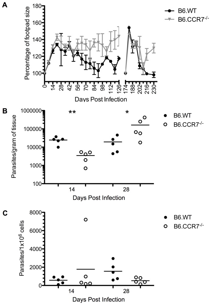
Figure 1. Disease progression in B6.CCR7-/- mice after infection with L. major.
(A) Mice were infected subcutaneously with L. major and the footpad lesion was measured weekly with a metric caliper. The lesion size was compared to the mean of the uninfected footpads from each experimental group (100%). Mice were infected subcutaneously in the contralateral footpad at day 174 post infection to measure the memory response. In accordance with animal ethics guidelines to minimise the usage of mice, this experiment was conducted at the same time as previously published data, and as a result, the lesion size of B6.WT mice has been published [61]. The parasite burdens in the footpad (B) and draining lymph node (C) were determined by limiting dilutions at days 14 and 28 post infection for B6.WT (closed circles) and B6.CCR7-/- (open circles) mice. One representative analysis of two experiments; Experimental group size: n=5 mice/genotype; *p<0.05; **p<0.01.

