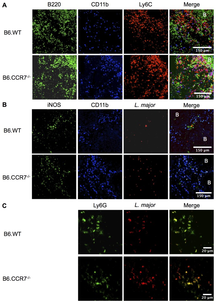
Figure 6. Fluorescent immunohistology of B6.WT and B6.CCR7-/- spleens at day 14 post infection and lymph nodes 24 hours after infection.
(A) Frozen spleens from day 14 post infection were sectioned and stained with antibodies against B220 (green), CD11b (blue) and Ly6C (red) to show localisation of monocytes in the spleen. (B) Expression of iNOS (green), CD11b (blue) and L. major (red) was shown in the spleen. (C) The draining popliteal lymph node was taken at 24 hours after infection with L. major expressing eGFP (red) and analysed for Ly6G (green) expression. B=B cell follicle; Objective magnification: A&B 20x; C 63x.

