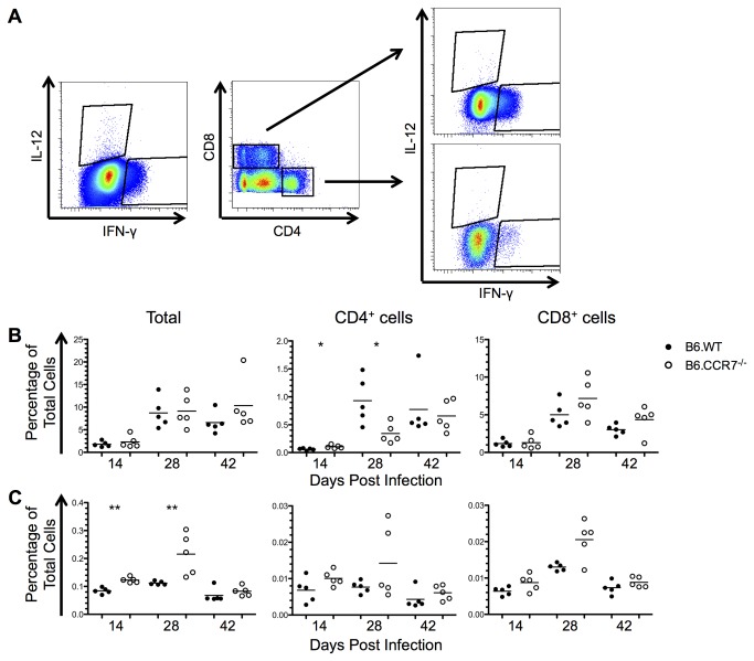
Figure 7. Cellular expression of Th1 cytokines in the lymph node of mice infected with L. major.
(A) Intracellular expression of IL-12 and IFN-γ was determined by flow cytometry. (B) The expression of IFN-γ in B6.WT (closed circles) and B6.CCR7-/- (open circles) mice is shown as a percentage of all lymph node cells as well as a the percentage of CD4+ or CD8+ cells expressing IFN-γ. (C) The expression of IL-12 was shown in a similar manner to IFN-γ. One representative analysis of three experiments; Experimental group size: n=5 mice/genotype for each timepoint; *p<0.05; **p<0.01.

