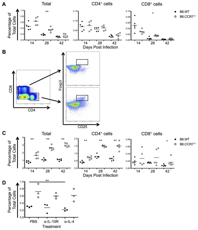
Figure 9. Immunosuppression in the lymph nodes of B6.WT and B6.CCR7-/- during infection with L. major.
(A) Expression of IL-10 in the total lymph node, as well as in the CD4+ and CD8+ compartments was determined by flow cytometry in B6.WT (closed circles) and B6.CCR7-/- (open circles) mice. (B) CD4+ or CD8+ Tregs were determined by their dual expression of CD25 and Foxp3. (C) The total percentage of Foxp3+ cells as well as the percentage of CD4+ and CD8+ Tregs in the total population was determined by intracellular flow cytometry. (D) Mice were injected intraperitoneally with PBS, anti-IL-10R or anti-IL-4, and the draining lymph node analysed for the percentage of CD4+ Tregs at day 14 post infection. A&C, One representative analysis of three experiments; Experimental group size: n=5 mice/genotype for each timepoint; D, One experimental analysis; Experimental group size: n=3 mice/genotype for each treatment. *p<0.05; **p<0.01.

