Figure 4.
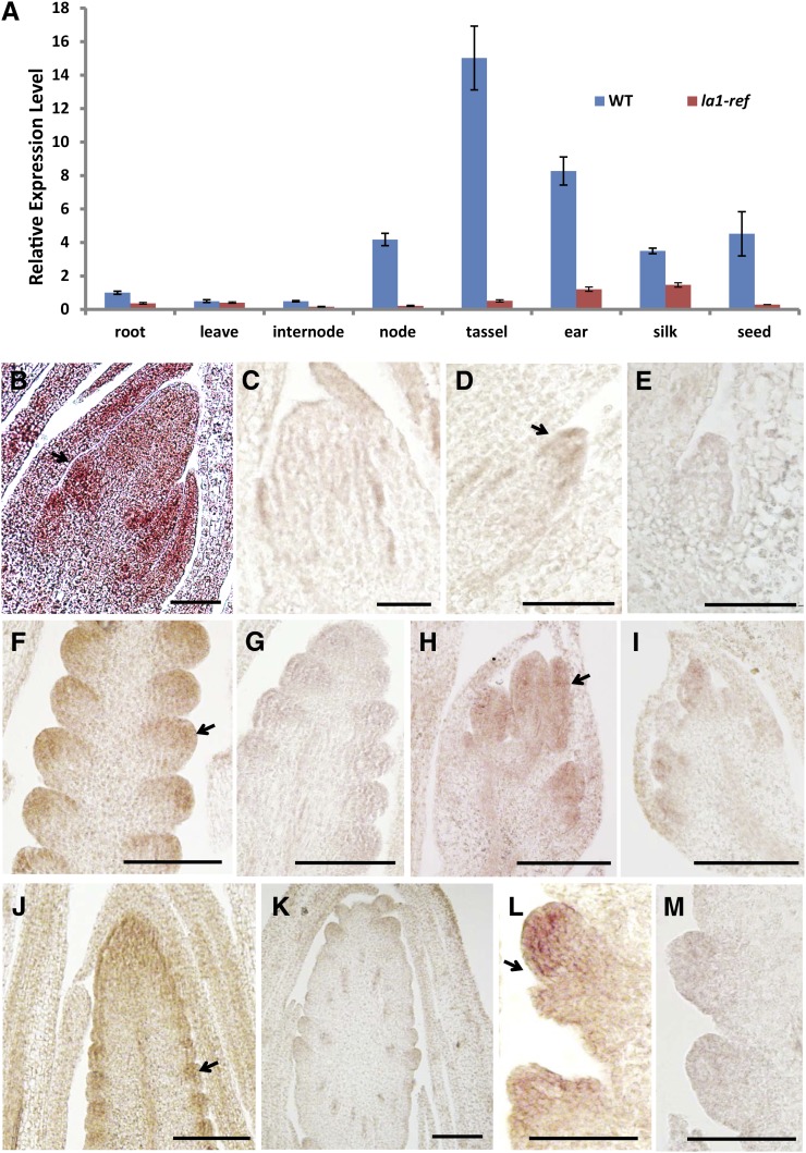
Expression pattern of ZmLA1. A, qRT-PCR analysis of ZmLA1 in multiple tissues in wild-type (WT) and la1-ref plants. Three biological replicates were performed for each sample, and Actin1 (GRMZM2G126010) was used as an internal reference. Error bars indicate the se between biological replicates. B to M, In situ hybridization of ZmLA1 in wild-type (B, D, F, H, J, and L) and la1-ref mutant (C, E, G, I, K, and M) plants. All samples are 10-µm longitudinal sections: 2-week-old shoot apical meristems (B and C); axillary meristem (D and E); developing SPMs on the flank of inflorescence meristem of 34-d-old tassels (F and G); developing male flower primordia (H and I); 54-d-old ears (J and K); and developing SPMs on the flank of inflorescence meristem of 54-d-old ears (L and M). Arrows indicate the expression of ZmLA1. Bars = 50 µm.

