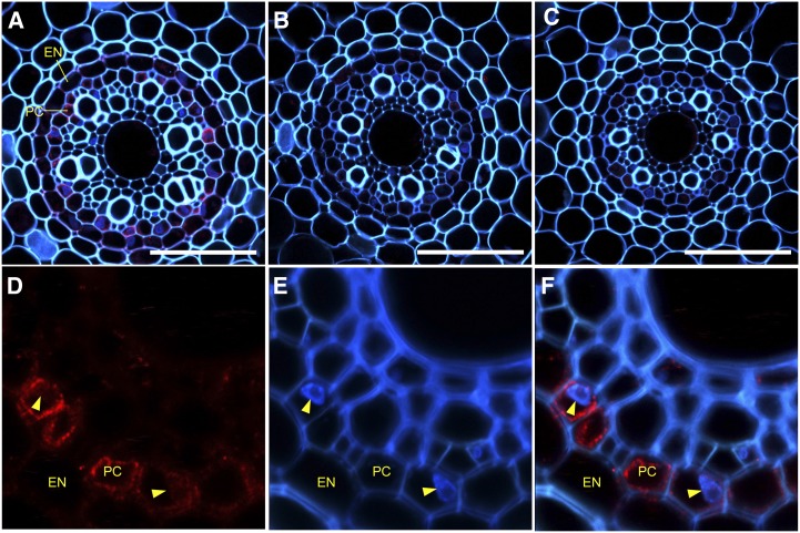
Figure 3.
Localization of OsHMA5 in rice roots. Immunohistochemical staining of OsHMA5 with anti-OsHMA5 polyclonal antibody was performed in the roots of wild-type rice (A and D–F) and two knockout lines, NE6050 (B) and NF8524 (C). Magnified images of the stele in the wild type are shown in D to F. The signal of anti-OsHMA5 antibody (D; red), autofluorescence of cell wall and nuclei stained by DAPI (E; blue), and the merged image (F) are shown. The nucleus is indicated by yellow arrowheads. EN, Endodermis; PC, pericycle. Bars = 50 µm.

