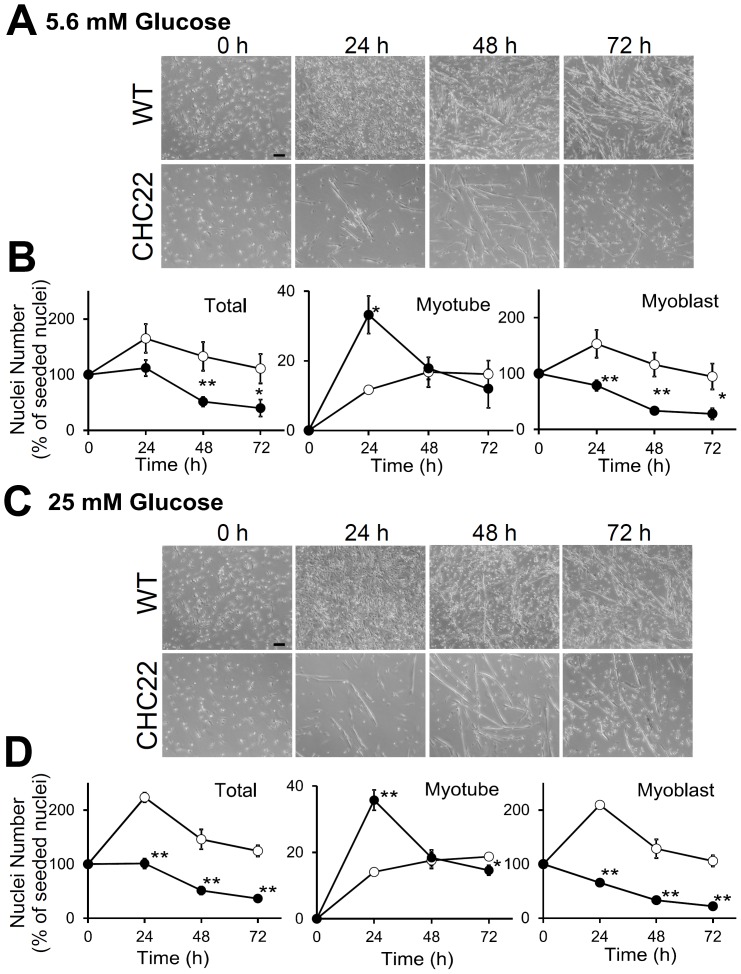
Figure 7. Myoblasts from CHC22-mice undergo fusion but do not exhibit glucose-dependent proliferation.
A and C) Images of primary myoblasts from wild-type (WT) or CHC22-transgenic mice cultured in FM with A) low (5.6 mM) or C) high (25 mM) glucose for the indicated time in hours (h), all seeded at the same density (scale bars, 100 µm). B and D) At the indicated time period for myoblasts cultured as in A and C, the nuclei were quantified for total number (Total), number present in multi-nuclear myotubes (Myotube), and number in mono-nuclear cells (Myoblast). These quantifications are plotted relative to the total nuclei present at the start of differentiation (switch to FM at 0 hours) for cultured myoblasts from CHC22-mice (filled circles) and WT mice (open circles). Two-way ANOVA and Tukey-Kramer post-hoc test showed significant differences between WT and CHC22 in all three panels. (*p<0.05 and **p<0.01) at indicated time points.

