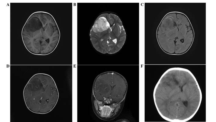
Figure 1.
EVN identified in the right frontal cortex and subcortical white matter of a 2-year-old female. (A) T1-weighted image, (B) T2-weighted image, (C) FLAIR image and (D) axial and (E) coronal post-contrast T1-weighted image showing a moderate-to-marked diffuse and enhanced right frontal lobe lesion involving mainly the cortex and subcortical white matter. Peritumoral edema, cysts, hemorrhages and calcification were not observed. (F) CT scan revealed a large, microcystic and non-calcified right frontal lobe mass. EVN, extraventricular neurocytoma; FLAIR, fluid-attenuated inversion recovery.

