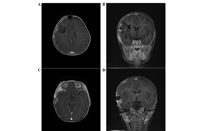
Figure 5.
MRI scan with enhancement performed 10 weeks after the initial resection, showing an enlargement of the two tumor nodes identified six weeks previously. (A and C) Axial and (B and D) coronal images were captured. Notably, the tumor node located in the initial surgery field showed mild enhancement, but the other tumor node located in the area posterior-inferior to the initial surgery field showed significant enhancement. MRI, magnetic resonance imaging.

