Figure 5:

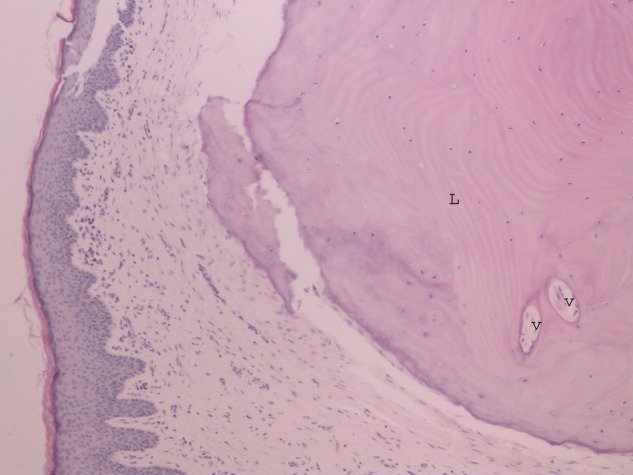
Photomicrograph of osteoma within the skin of external auditory canal showing lamellar bone (L) and fibrovascular channel (V) (H&E ×20).

Photomicrograph of osteoma within the skin of external auditory canal showing lamellar bone (L) and fibrovascular channel (V) (H&E ×20).