Figure 1.

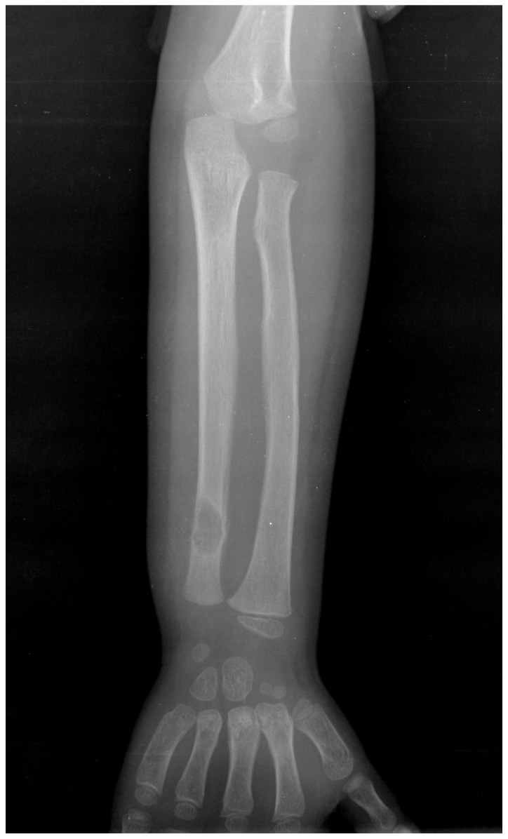
Case 1. X-ray demonstrating a well-circumscribed osteolytic lesion with slight marginal sclerosis involving the diaphysis of the distal left ulna.

Case 1. X-ray demonstrating a well-circumscribed osteolytic lesion with slight marginal sclerosis involving the diaphysis of the distal left ulna.