Figure 1.
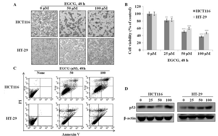
Effects of epigallocatechin-3-gallate (EGCG) on proliferation and apoptosis in HCT116 (wild-type p53) and HT-29 (mutant p53) colon cancer cells. (A) HCT116 and HT-29 cells were treated with or without various concentrations of EGCG for 48 h, and cell morphology was examined by light microscopy (×400). (B) Cell viability was determined by 3-(4,5-dimethylthiazol-2-yl)-2,5-diphenyltetrazolium bromide (MTT) assay, and is represented as the percentage of relative absorbance compared to controls. (C) Representative flow cytometric data from HCT116 and HT-29 cells that were treated with or without EGCG for 48 h and then stained with Annexin V and PI. (D) Cells were treated with various concentrations (25–100 μM) of EGCG for 24 h, and p53 protein expression was analyzed by western blotting. **P<0.01 and ***P<0.001 vs. control (0 mM).

