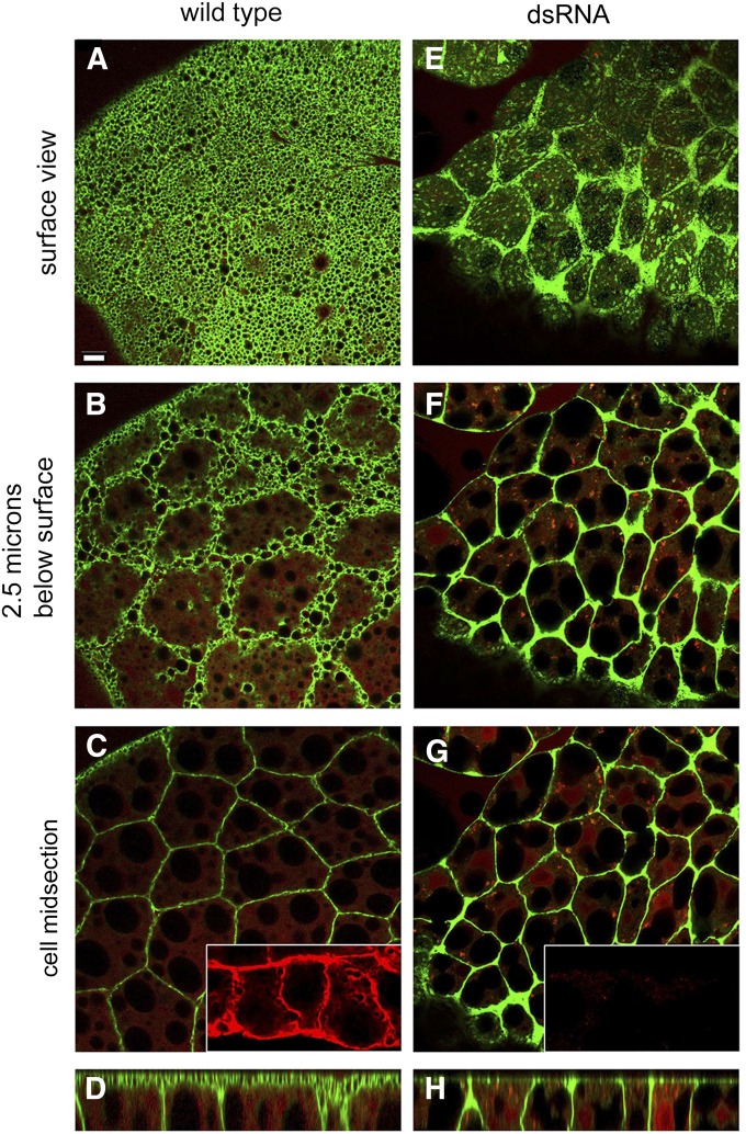
Figure 1.
The foamy appearance of the larval fat body surface was lost with knockdown of β-spectrin. (A) The plasma membrane marker mCD8-GFP (green) produced an unusual foamy pattern of surface labeling in the wild-type larval fat body. (B and C) Deeper confocal sections revealed a more typical polygonal profile of cell–cell contacts. (E–G) After β-spectrin knockdown with RNAi the foamy appearance was replaced by a coarse speckled appearance, although the polygonal pattern at cell contacts remained. The β-spectrin knockdown was efficient as judged by anti-β-spectrin staining (C and G, insets, red). (D and H) A rotated view (compiled from a Z series using ImageJ) revealed a broad zone of ecto domain mCD8-GFP fluorescence in the wild type (D) that was lost after β-spectrin knockdown (H). Cells were counterstained by cytoplasmic DSRed expression, which outlines the large lipid droplets present in each cell. Bar, 10 µm.

