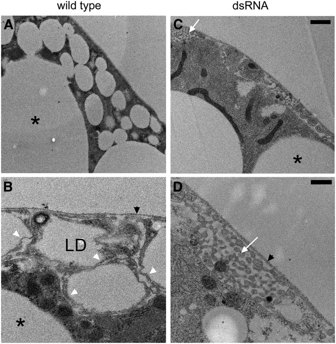
Figure 2.
Electron microscopy revealed that a population of small, cortical lipid droplets in larval fat body explains the foamy appearance with fluorescent markers. (A and B) In wild type, small lipid droplets (LD) were found closely apposed to the plasma membrane. The small cortical lipid droplets (1–4 μm) were distinct from the much larger lipid droplets (*) found deeper in the cytoplasm. (C and D) The population of small cortical LD was absent after knockdown of β-spectrin. Higher magnification views revealed that the cortical lipid droplets were housed within small protuberances of the plasma membrane, tightly packed between the prominent extracellular matrix that surrounds the fat body (black arrowheads) and the rest of the cell. White arrowheads mark the thin rim of cytoplasm found between LD and the plasma membrane. (D) The lipid droplets and protuberances were no longer visible after β-spectrin knockdown. Instead there was a peculiar pattern of smaller, interconnected tubular structures (white arrows) in pockets formed between the extracellular matrix and the cell body. Bars, A and C, 1 µm; B and D, 0.5 µm.

