Figure 3.
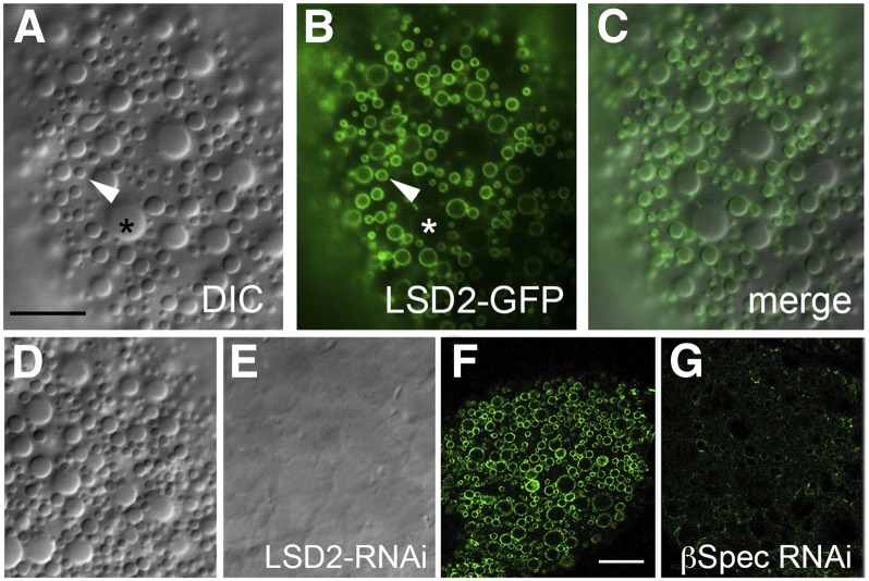
The small cortical lipid droplets are Lsd-2 positive. (A,D) Lipid droplets in the ecto domain of fat body cells can be detected by DIC microscopy. (B and F) Expression of UAS‐Lsd-2-GFP in the fat body (via Cg-Gal4) produces a pattern of small lipid droplets labeled on their surface (B) that exactly coincides with the DIC pattern (merge in C). Larger lipid droplets (>4 μm) were not labeled by Lsd-2‐GFP (B, *). (E) Knockdown of Lsd-2 by RNAi resulted in disappearance of the cortical lipid droplets. (G) Likewise, knockdown of β-spectrin by RNAi eliminated the population of Lsd‐2‐GFP-labeled vesicles in the cortex. Bars, A–E, 10 μm; F and G, 20 μm.

