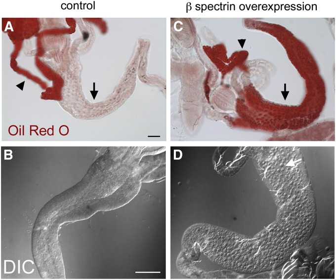
Figure 6.
Oil Red O staining of lipid droplets in dissected preparations of wild-type and β-spectrin-overexpressing larvae. (A) Most of the Oil Red O staining in wild type was confined to the fat body (arrowhead) with only a trace of staining visible in the midgut epithelium (arrow). (C) There was a dramatic increase in anterior midgut staining in larvae that overexpressed UAS‐β‐Spec95 at 25°. (B and D) The change was also visible by DIC: lipid droplets were rarely detectable in controls (B) and there was a dramatic increase in lipid droplets in the anterior midgut (white arrow) upon UAS‐β-Spec95 overexpression (D). Bars, A and C, 50 µm; B and D, 20 µm.

