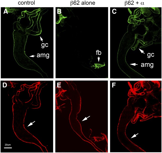
Figure 9.
Overexpression of β-spectrin alters the behavior of lipophorin. (A–F) Dissected larvae expressing UAS‐β‐Spec62 alone (B and E) or together with UAS‐α-Spec37 (C and F, as in Figure 8) in the fat body (Cg‐Gal4) were double labeled with anti‐lipophorin antibody and FITC secondary antibody (A–C, green) and anti-β-spectrin antibody and TR secondary antibody as a staining control (D–F, red). Lipophorin antibody stained the outer surface of the anterior midgut (amg) and gastric caeca (gc) of wild-type larvae (A). Midgut lipophorin staining was absent when β-spectrin was overexpressed in the fat body (B), although it was still abundantly detected within the fat body (fb). Lipophorin staining of the midgut was restored in rescued larvae expressing both α- and β-spectrin transgenes (C). Bar, 20 µm.

