Figure 9.
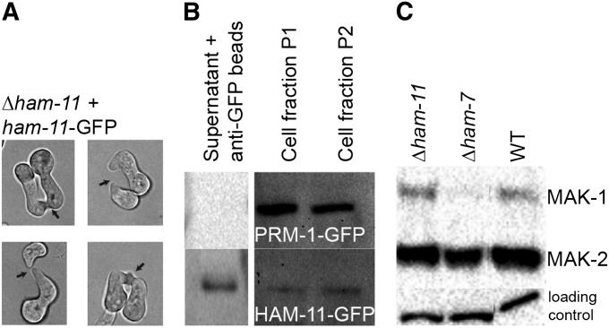
MAK1 and MAK2 phosphorylation, fusion phenotype, and Western blot of Δham-11 complemented with ham-11-gfp. (A) Four germling pairs of Δham-11 ham-11-GFP show chemotrophic growth and cell fusion (see arrows), showing full complementation of the Δham-11 phenotype by the ham-11-gfp construct. (B) Western blot of PRM1-GFP (which localizes to endomembranes and the plasma membrane of N. crassa by fluorescence microscopy) (Fleissner et al. 2009a). HAM11-GFP was detected in supernatant, endomembranes (cell fraction P1), and outer membranes (cell fraction P2). Cell fractionation purity was also assessed using antibodies to the plasma membrane ATPase (data not shown), which localized only to the membrane fractions. (C) Activation of MAK1 and MAK2 in wild type, Δham-11, and Δham-7 germlings. Protein samples from Δham-11 (lane 1), Δham-7 (lane 2), and wild-type (lane 3) 5-hr-old germlings from liquid VMM are shown. Phosphorylated MAK1 (51 kDa) and MAK2 (41 kDa) were detected using anti-phospho p44/42 MAP kinase antibodies (Cell Signaling Technology). (Bottom) Shows equal loading for each lane.

