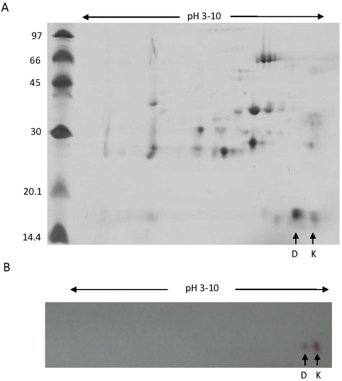
Figure 3.
2-DE SDS-PAGE pattern of venom proteins from B. leucurus. (A) 60 µg of total proteins were isoelectrically focused (pI range 3–10) followed by separation by SDS-PAGE (15% gel) and Coomassie blue staining. Protein spots of approx. 14 kDa correspond to the position of Bl-PLA2 isoforms are indicated by arrows, (B) Immunoblotting of venom proteins separated by 2D-SDS-PAGE with anti-BlK-PLA2 IgG. Arrows indicates the reactivity of the antibody with the approx. 14 kDa PLA2s.

