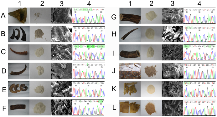
Figure 2. Sample information.
(A): black rhinoceros; (B): Asian water buffalo; (C): Mongolian gazelle; (D): domestic goat; (E): sheep; (F): red deer; (G): sika deer; (H): Père David's deer; (I): Chinese muntjak; (J): Hawksbill; (K): Chinese softshell turtle; (L): Reeve's turtle. 1: Photograph of animal organs; 2: powdered animal organs, which represent the main market form and which are hard to discriminate; 3: a scanning electron microscope photograph of animal organs (obtained using a JSM-6510 instrument, Japan; gold sputtering treatment: 40 mA, 130 s; SEI: 12 kV; WD: 12 mm; amplification × 5500), no typical diagnostic characteristics are found; 4: sequencing peaks (the peaks were clipped using Codoncode Aligner, and the 24-bp segment can be used to identify all of the animal organ samples). (The photographs in this figure were taken by JL, and we hold the copyright of this figure).

