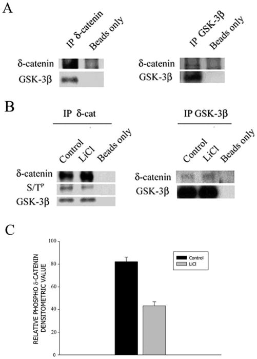
Figure 2. δ-Catenin co-immunoprecipitates with GSK-3β in neuronal tissues.
A. (Left panel) Western blot analysis of δ-catenin immunoprecipitated (IP) from mouse brain homogenates and probed with indicated antibodies; (Right panel) GSK-3β IP from mouse brain homogenates probed with indicated antibody. B. Primary cortical neurons were treated with GSK-3β inhibitor (LiCl) as indicated and immunoprecipitated. (Left panel) δ-catenin IP probed with GSK-3β and phospho-serine threonine antibody (S/TP); (Right panel) GSK-3β IP probed with indicated antibodies. C. Bar graph shows mean values of relative densitometric phospho-δ-catenin immunoreactivity. Error bars represent ± SEM with n =3.

