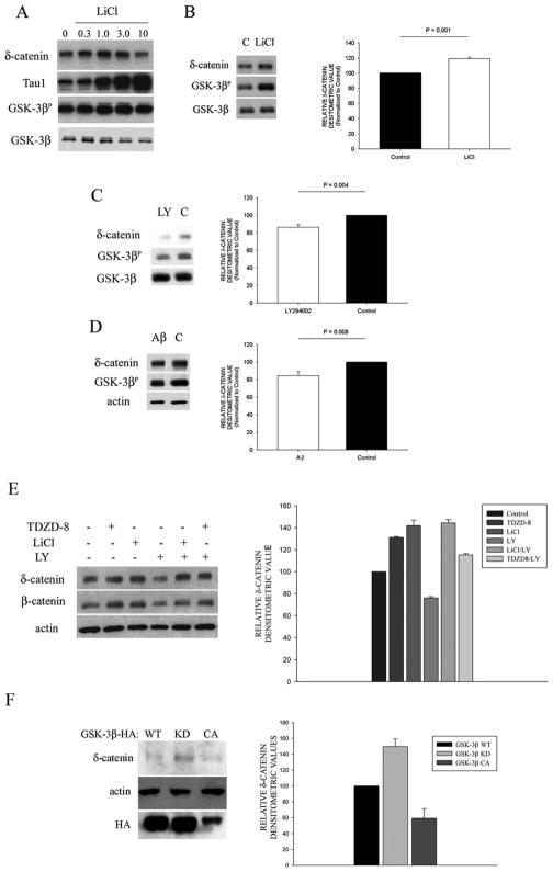
Figure 3. Regulation of δ-catenin by the GSK-3β pathway in primary cortical neurons.
A. Western blot analysis showed that inhibition of GSK-3β with LiCl (up to 1.0 mM) increased δ-catenin levels in primary cortical neurons. Tau1 (dephospho-Tau) was used to verify effectiveness of pharmacological treatment. GSK-3β activity was verified by probing with GSK-3βP-Ser9 (numbers represent mM concentrations). B. Western blot analysis of primary cortical neurons treated with 1 mM of LiCl, shows decreased δ-catenin expression compared with untreated controls (C). C. Inhibition of PI3K activity with LY294002 (LY) decreased δ-catenin protein expression in primary cortical neurons. D. Treatment of neurons with Aβ42 (Aβ) decreased the amount of phosphorylated GSK-3β and showed decreased δ-catenin expression. E. Neurons treated with LiCl, as well as a different GSK-3β specific inhibitor TDZD-8 (GSK-3βi), showed increased δ-catenin and β-catenin immunoreactivity. Four-hour pretreatment of neurons with GSK-3β inhibitors shows rescue of δ-catenin and β-catenin expression in the presence of LY294002. F. CWR22-Rv-1 cells transfected with either GSK-3β-HA WT (wild type), GSK-3β KD (kinase dead), and GSK-3β CA (constitutively active). Transfection with GSK-3β KD and GSK-3β CA demonstrate corresponding increases and decreases in endogenous δ-catenin immunoreactivity. Bar graphs to the right of representative Western blots in panel B, C, D, E, and F show mean values of relative densitometric of δ-catenin from Western blots. Analysis was performed with SigmaStat Version 3.11 (Systat Software, Inc., San Jose, CA) using the t-test module. Error bars represent ± SEM with n = 5 (LiCl); 4 = (LY); 4 = (Aβ); 3 = (E and F).

