
Figure 5. Localization of δ-catenin with GSK-3β destruction complex members in the neurites of primary hippocampal neurons.
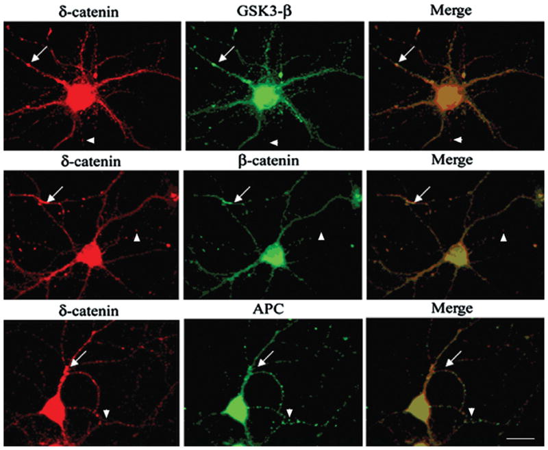
Double immunofluorescence staining shows intracellular distribution of δ-catenin, β-catenin, GSK-3β, and APC. Arrows show co-localization of GSK-3β destruction complex molecules with δ-catenin whereas arrowheads show non-overlapping localization of δ-catenin with β-catenin, GSK-3β, and APC. Bar: 15 μm.