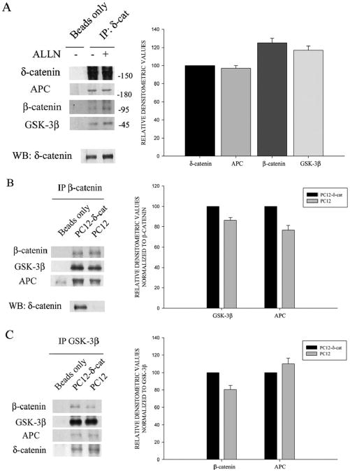
Figure 6. δ-Catenin expression facilitates β-catenin, GSK-3β, and APC interactions.
A. δ-Catenin co-immunoprecipitated with APC, β-catenin, and GSK-3β from cultured neurons. ALLN treatment showed an enhanced co-immunoprecipitation of δ-catenin with β-catenin and GSK-3β. Bar graph to the right represents mean densitometric values from δ-catenin immunoprecipitated cells treated with ALLN. Densitometric values for APC, β-catenin, and GSK-3β were compared to δ-catenin. B. and C. Lysates from tetracycline-inducible PC12 cells overexpressing δ-catenin (PC12-δ-cat) were immunoprecipitated with either anti-β-catenin or anti-GSK-3β antibodies as indicated. Immunoprecipitated samples were then immunoblotted with indicated antibodies. Greater amounts of GSK-3β (B) and β-catenin (C) were co-precipitated from PC12-δ-cat cells compared to PC12. Non-immunoprecipitated samples were subject to Western (WB) analysis (bottom panel) using anti-δ-catenin antibody. Bar graphs to the right of B and C demonstrate Western blot densitometric values of β-catenin, GSK-3β and APC from PC-12-δ-cat and PC12 cells. Data shown in panels A, B, and C are representative of gels from three separate experiments.

