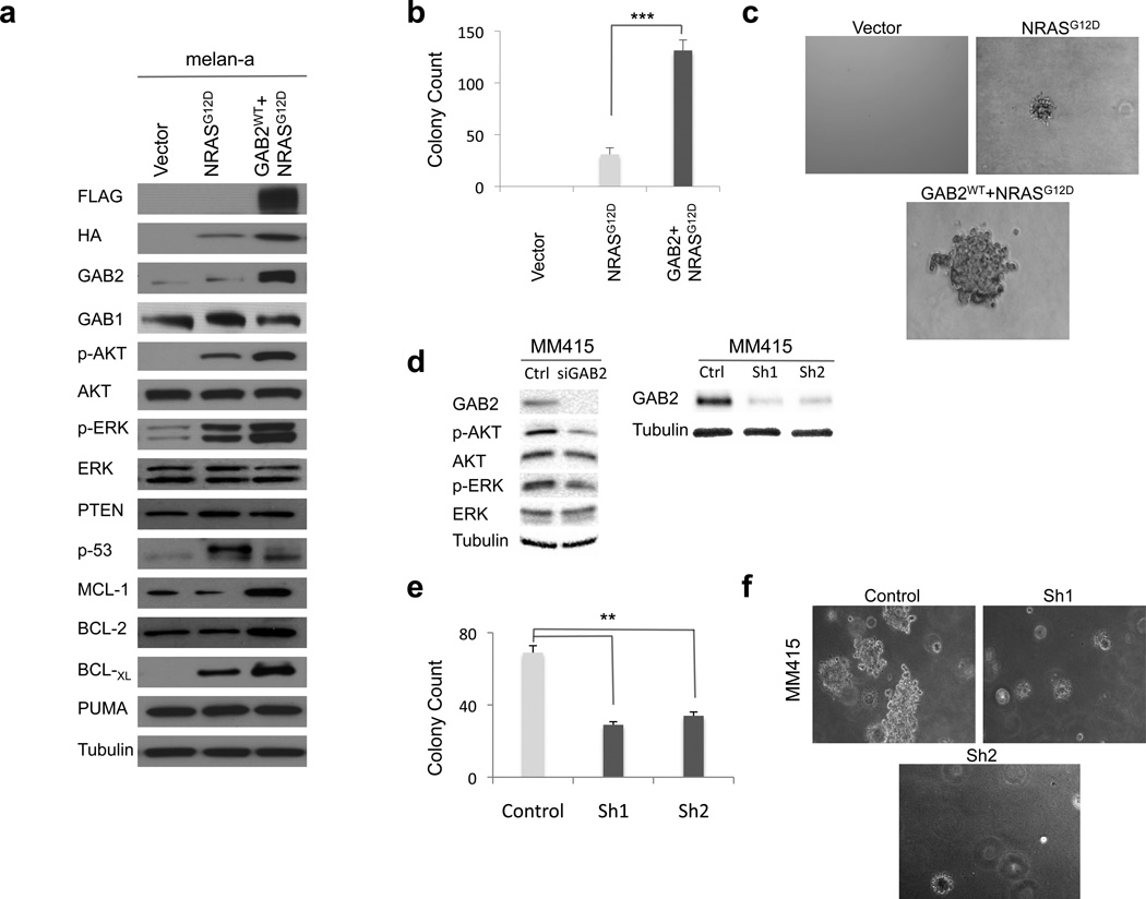
Figure 3. Co-expression of GAB2WT with NRASG12D promotes anchorage independence in melanocytes.
(a) Melanocytes stably expressing empty vector, NRASG12D (HA-tagged), or GAB2WT+NRASG12D (FLAG- and HA-tagged, respectively) were cultured and protein lysates were assayed by Western blotting. PI3K and MAPK activation were examined by measuring phospho-AKT and phospho-ERK levels, respectively. Expression of PUMA, p53 and BCL-2 family of anti-apoptotic proteins, MCL-1, BCL-2 and BCL-XL, were also evaluated. Tubulin was used as loading control.
(b) and (c) Quantification and representative images of soft agar colony formation assay of melanocytes expressing vector, NRASG12D, and GAB2WT+NRASG12D are shown. Briefly, 1×10⁵ cells were resuspended in 0.35% agarose and overlaid onto 0.7% agarose in growth medium containing 10% FBS in 6-well plates. Colonies were stained with thiazolyl blue tetrazolium bromide and counted at 4 weeks after plating. Experiments were performed in triplicates. Error bars indicate ± standard error. *p<0.05, **p<0.01, and ***p<0.001.
(d–f). MM415 is a mutant NRAS melanoma cell line with GAB2 overexpression. GAB2 was silenced with scrambled (control) or siRNA and GAB2-dependent signal transduction pathways were analyzed by western blotting. Stable knockdown of GAB2 was achieved by infecting cells with control, Sh1 and Sh2. Quantification and representative images of soft agar colony formation assay of melanomas expressing control and shGAB2 are shown. Colonies were counted two weeks after plating. Experiments were performed in triplicates. Error bars indicate ± standard error. *p<0.05, **p<0.01.

