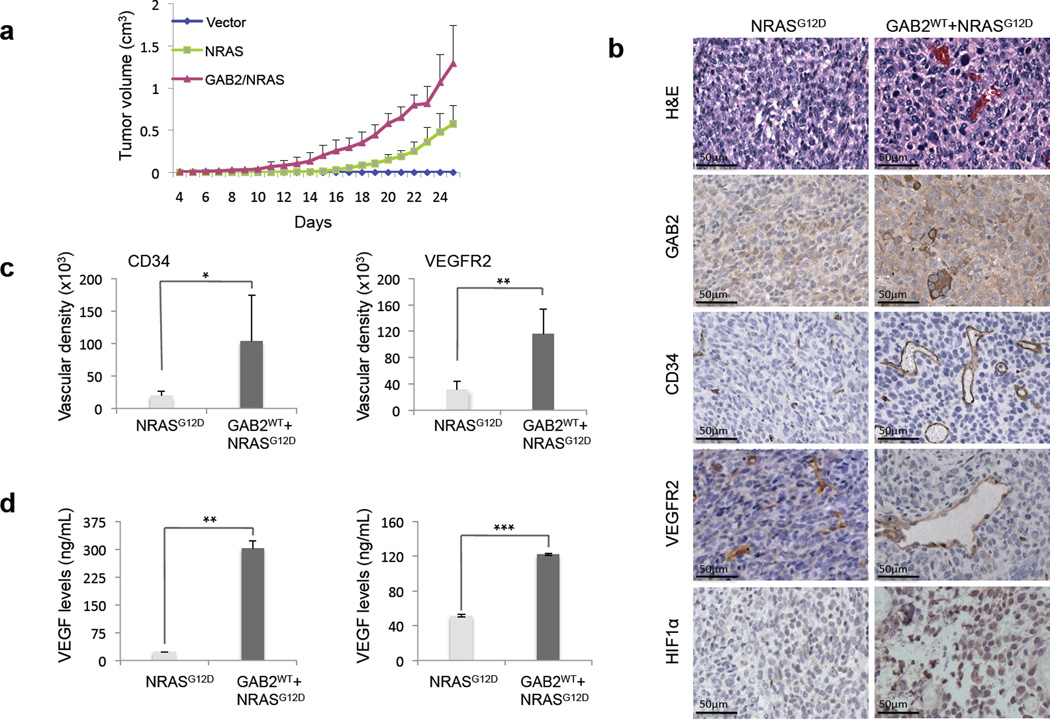
Figure 5. GAB2 enhances tumorigenesis and induces angiogenesis in vivo.
(a). 1×10⁶ melan-a melanocytes stably expressing vector, NRASG12D, GAB2WT+NRASG12D were injected into nude mice subcutaneously (n=8 mice per group). Primary tumor growth was examined by measuring tumor volume every other day.
(b). At four weeks, xenograft tumors were analyzed histologically using hematoxylin and eosin staining as well as for GAB2, CD34, VEGFR2, and HIF1α, using immunohistochemistry. Representative micrographs of NRASG12D and NRASG12D+GAB2WT expressing tumors are shown.
(c). CD34 or VEGFR2-staining vasculature was quantified by measuring the vessel areas (20× per field, 4 fields per section) using the Image J software. Error bars indicate ± standard error. *p<0.05, **p<0.01.
(d). Proteins were extracted from xenografts (left panel) or cells cultured in 2D conditions (right panel) expressing either NRASG12D or GAB2WT+NRASG12D, and VEGF levels were measured by ELISA. Experiments were performed in duplicates. Error bars indicate ± standard error. **p<0.01, ***p,0.001.

