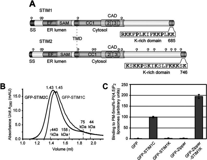
Figure 1. Tetramerization of STIM1 K-rich domain leads to PI(4,5)P2 binding.
(A) Schematic outline of human STIM1 and STIM2 domain structure (drawn to scale). SS (signal sequence), EF-hand domain (EF), SAM, TMD (transmembrane domain), CC domains 1–3, CAD, the K-rich domains with their amino acid sequences and the orientations of N- and C-termini are shown. Cysteine residues are depicted as grey circles. (B) Purified GFP-tagged CTDs of STIM1 and STIM2 were run on S200 gel-filtration column and the elution profiles of GFP–STIM1C (grey), GFP–STIM2C (black) and of the indicated marker proteins are shown. (C) Binding of 1 μM of each GFP, GFP–STIM1C, GFP–STIM1K, GFP-tagged GCN4 leucine zipper (GFP–Zipper) and GFP–Zipper with STIM1 K-rich domain (GFP–Zipper–STIM1K) to PM-like liposomes with 5 mol% PI(4,5)P2 in presence of 1 mM DTT. Binding of GFP–STIM1C was set to 100 and bars indicate mean±S.D. from at least three experiments.

