
Figure 1.
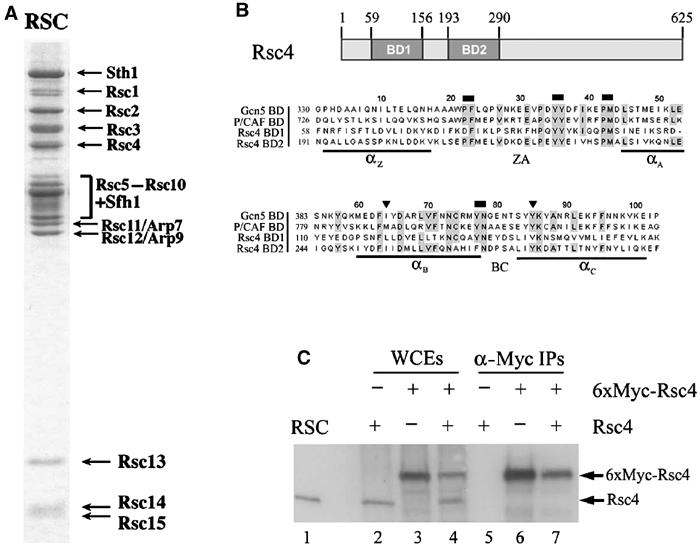
Identification of Rsc4. (A) Purified RSC complex stained with Coomassie dye (from Cairns et al, 1996). (B) Domain structure of Rsc4 and alignment of Rsc4 bromodomains with the bromodomains of yeast Gcn5 and P/CAF. Regions of identity are highlighted in gray. Triangles (▾) mark residues mutated in rsc4-2. Rectangles (⁃) mark paired residues mutated by site directed mutagenesis. (C) Stoichiometry of Rsc4 in RSC. Western analysis using anti-Rsc4 antiserum of whole-cell extracts (WCEs) and anti-Myc precipitations from three strains bearing different tagged RSC4 alleles. Extracts were prepared from YBC627 transformed with either 6xMyc-Rsc4 (p603) (lanes 3, 4, 6, and 7) or the empty vector (p415.MET25, p520) (lanes 2 and 5). Cells used in lanes 3 and 6 lost the untagged Rsc4 plasmid on SC+5-FOA.