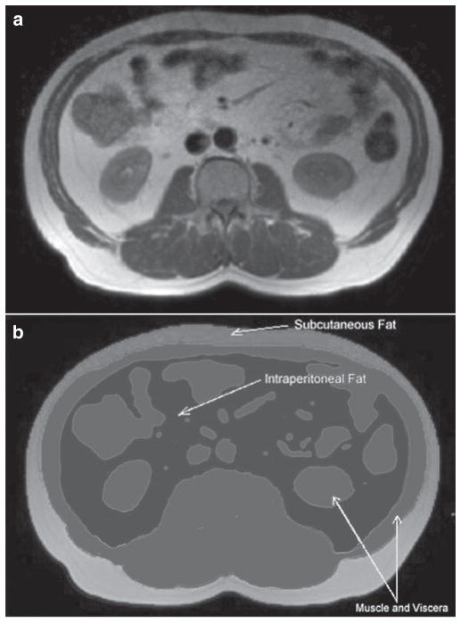
Figure 2.
Identification of fat depots. (a) An axial slice used for quantitation of fat and (b) highlights color shadowing used to segment the abdominal slice into visceral organs and fat depots. Voxels identified in white represent subcutaneous fat, those in black demarcate intraperitoneal fat. Gray voxels identify muscular tissue and viscera.

