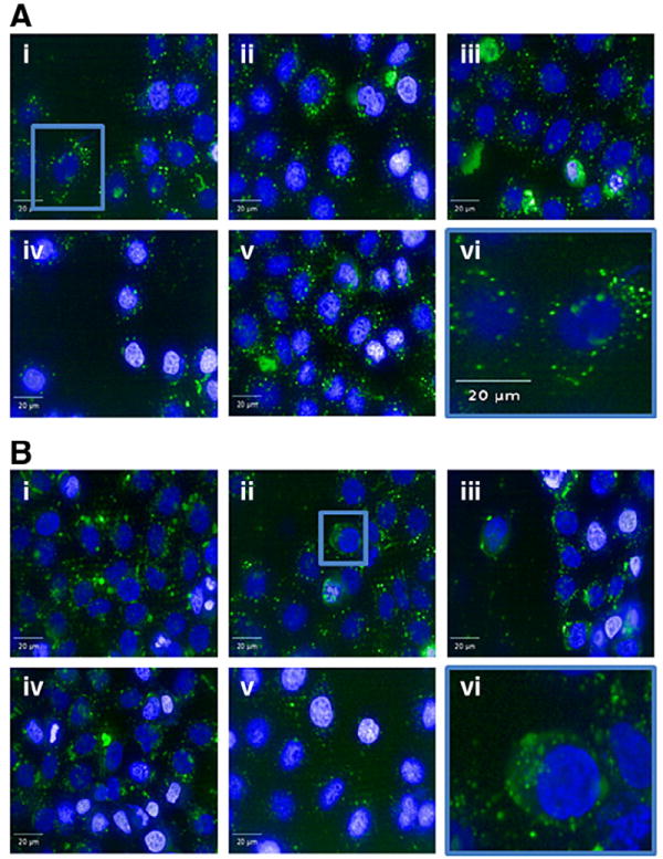
Fig. 4.

Cellular uptake of lipid–PEI–siRNA complexes. The uptake of lipid–PEI compounds complexed with Cy5-labled siRNA by HeLa cells was imaged using an Opera spinning disk confocal microscope. The uptake of the lipid:PEI mole ratio of 0.56 (A) is compared to that of lipid:PEI mole ratio of 2.91 (B). Five different representative images are presented for each compound (i–v) and image (vi) represents an enlarged image. Punctate structures can be noticed in panel A (vi), while cytoplasmic as well as punctuate structures can be seen in panel B (vi). Blue is Hoechst dye.
