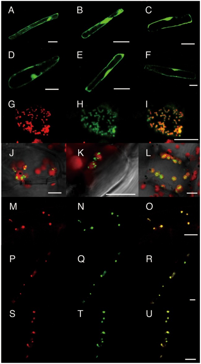
Fig. 5.

Subcellular localization of GFP-tagged OsIPT proteins in Arabidopsis observed by confocal laser-scanning microscopy. The translational fusion genes OsIPT1-GFP (A), OsIPT2-GFP (B), OsIPT3-GFP (C), GFP-OsIPT1 (D), GFP-OsIPT2 (E) and GFP-OsIPT3 (F) were transiently expressed in root epidermal cells by particle bombardment. The fusion genes pGGPS6-DsRed2 (G), a control for mitochondrial localization, and OsIPT7-GFP (H) were co-introduced into a leaf mesophyll cell. (I) Merged image of (G) and (H). The fusion genes OsIPT4-GFP (J), OsIPT5-GFP (K) and OsIPT8-GFP (L) were introduced into leaf mesophyll cells and superimposed on Chl autofluorescence (red). The fusion gene AtFSD3-DsRed2 (M, P and S), a control for nucleoid localization, was co-introduced into root epidermal cells with either OsIPT4-GFP (N), OsIPT5-GFP (Q) or OsIPT8-GFP (T). (O, R and U) Merged images of (M) and (N), (P) and (Q), and (S) and (T), respectively. Scale bars, 20 µm (A–F) and 10 µm (G–U).
