
Figure 5.
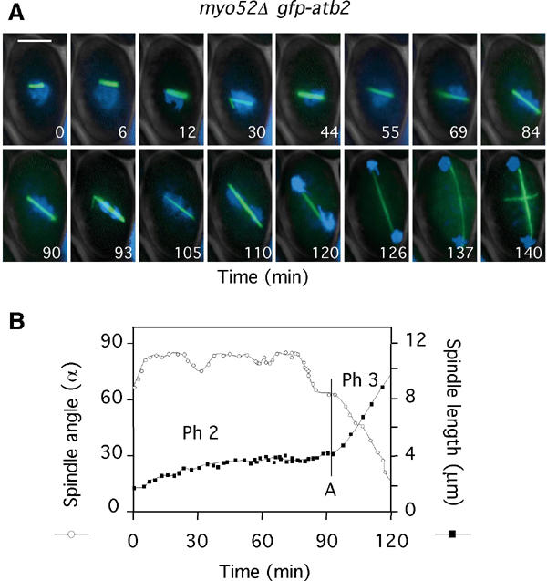
The spindle orientation checkpoint is activated by disruption of the CAR. (A) myo52Δ gfp-atb2 cells were filmed in medium containing Hoechst (Supplementary Movie 7). Numbers indicate the time from when the spindle attained 2 μm. Bar=4 μm. (B) Analysis of spindle angle (open circles) and spindle length (closed squares) in myo52Δ gfp-atb2 cells (Supplementary Movie 7). The vertical bar denotes the time of chromosome separation (anaphase A).