
Figure 6.
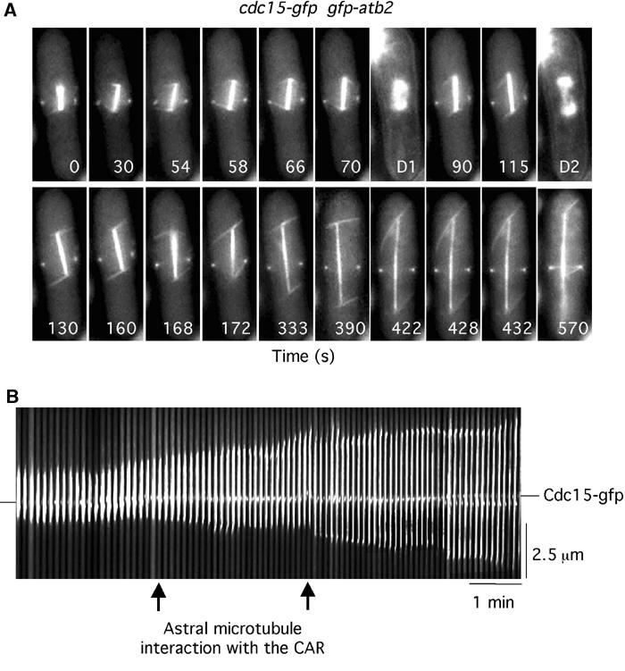
Astral microtubules interact with the CAR. (A) cdc15-gfp gfp-atb2 cells were filmed in medium containing Hoechst. Numbers indicate the time from the beginning of the movie (Supplementary Movie 8). D1 and D2 are images of chromatin staining. (B) Kymographic analysis of spindle position relative to the CAR in cdc15-gfp gfp-atb2 cells. In all, 100 images are shown (each 4 pixels wide). Arrows indicate the point of interaction of the astral microtubule from the lower SPB with the cortex.