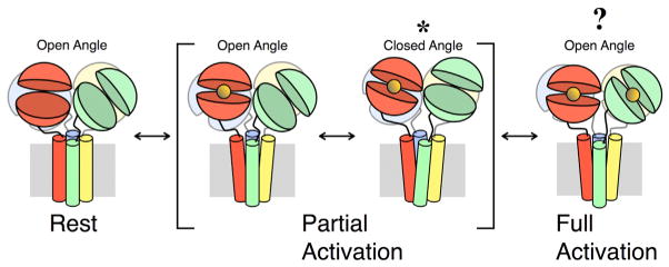
Figure 7. Proposed Structural Transitions Involved in AMPA Receptor Activation.

Subunits A, B, C, and D are colored green, red, blue, and yellow, respectively. The LBDs are arranged in the OA conformation when the receptor is in a state of rest. When the LBDs of subunits B and D bind glutamate (orange sphere), the receptor enters a state of partial activation. In this state, the LBDs may adopt either the OA or CA conformation. The conformation trapped by either the disulfide bridge formed by the A665C mutation or the metal bridge formed by one of the triple-residue mutations is indicated by an asterisk. When glutamate is bound to all LBDs, the receptor enters a state of full activation. We have no information about the LBD conformation in this state. It is unknown whether the ATD layer undergoes large-scale structural rearrangements during gating transitions, and it is therefore omitted from this figure. See also Figure S7 and Table S1.
