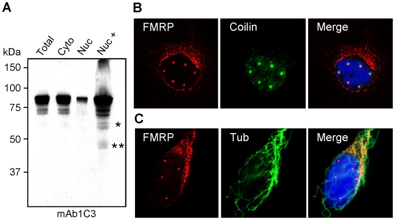
Figure 2. FMRP is present in the isolated nuclear fraction but not in nuclei.
(A) Total, cytoplasmic, and nuclear cytoplasmic fractions from HeLa cells were loaded in equal ratios as well as one overloaded nuclear fraction and analyzed by immunoblotting with mAb1C3 to determine the distribution of FMRP. Nuc+ refers to concentrated (20 µg) nuclear protein. (B) Double immunofluorescent localization of FMRP with IgYC10 (red) and Coilin (green) after gentle lysis of the cells in situ. Nuclei were counterstained with DAPI. (C) Double immunofluorescent staining of FMRP with IgYC10 (red) and cold-resistant microtubule network revealed with an anti-tubulin antibody (green). Nuclei were counterstained with DAPI. Due to the three dimensional distribution of microtubules, images were taken by conventional epifluorescent microscopy to reveal the microtubule framework.

