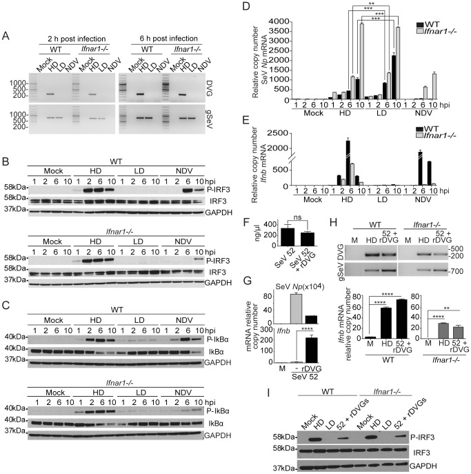
Figure 1. Robust and sustained activation of IRF3 and NF-κB independently of type I IFN feedback in response to SeV DVGs.
WT and Ifnar1−/− BMDCs were infected with SeV Cantell HD (HD), SeV Cantell LD (LD), or NDV (moi = 1.5 TCID50/cell). (A) Total RNA was extracted at 2 and 6 h post-infection and analyzed for the presence of DVGs and standard virus genome (gSeV) by PCR. Position of base pair size reference markers is indicated in each gel. Whole cellular extract was prepared and examined for phosphorylation of (B) IRF3 and (C) IκBα by western blot at the indicated time points. Total RNA was extracted from the infected cells and the expression of (D) SeV Np or NDV Hn mRNA and (E) Ifnb was monitored by RT-qPCR. (F) Total RNA content on equivalent infectious doses of SeV strain 52 or SeV 52 containing rDVGs. (G) LLC-MK2 cells infected with a moi = 5 TCID50/cell SeV 52 alone or in the presence or a recombinant (r)DVG. Expression of viral Np and Ifnb mRNA was determined by RT-qPCR 18 h after infection. (H) WT and Ifnar1 −/− mouse embryo fibroblasts were infected as indicated. Presence of WT or recombinant (r)DVG in the infected cells was determined by PCR 6 h after infection. Expression of the viral Np and Ifnb mRNA was determined by RT-qPCR 6 h after infection. (I) Whole cell extracts were analyzed for P-IRF3 by western blot. IRF3 and GAPDH are shown as controls. Gene expression is shown as copy number relative to the housekeeping genes Tuba1b and Rps11. Error bars indicate the standard deviation of triplicate measurements in a representative experiment (**p<0.01, ***p<0.001, ****p<0.001 by one-way ANOVA with Bonferroni post hoc test).

