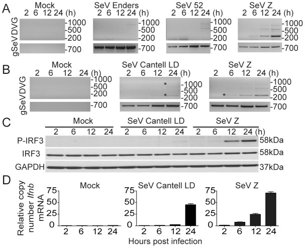
Figure 3. SeV copy-back DVGs generated in situ during infection have a strong stimulatory ability.
(A) TC-1 cells were either mock-infected or infected with a moi of 1.5 TCID50/cell of SeV strains Enders, 52, or Z. Total RNA was extracted at 2, 6, 12, and 24 h post-infection and analyzed for the presence of DVGs and standard virus genome (gSeV) by PCR. (B) BMDCs were infected with SeV strain Z or SeV Cantell LD and RNA was extracted at 2, 6, 12, and 24 h post-infection and analyzed for the presence of DVGs and gSeV. (C) Immunoblot of phosphorylated IRF3 in whole cell extracts from infected BMDCs and (D) Ifnb mRNA expression in infected BMDCs as determined by RT-qPCR. Gene expression is shown as copy number relative to the housekeeping genes Tuba1b and Rps11. Sequences from bands labeled with a star can be found in Fig. S5. Position of base pair size reference markers is indicated in each gel.

