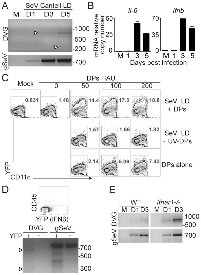
Figure 5. SeV copy-back DVGs are generated in the lung during infection.

(A) WT mice were infected with 104 TCID50/mouse of SeV Cantell LD or mock infected (M) and total RNA from lungs was analyzed for copy-back DVGs and standard viral genomes (gSeV) by PCR. (B) Expression of Ifnb mRNA in whole lung homogenates was analyzed by RT-qPCR. Gene expression is shown as copy number relative to the housekeeping genes Tuba1b and Rps11. (C) BMDCs from IFNβ-YFP mice were infected with a moi of 1.5 TCID50/cell of SeV Cantell LD alone, in the presence of purified DPs or UV-inactivated DPs, or with DPs alone and analyzed by flow cytometry 6 h post-infection. (D) CD45− cells were isolated from IFNβ-YFP mice 3 days after infection with SeV Cantell LD and sorted based on YFP expression. Total RNA from the YFP positive and negative fractions was extracted and analyzed for copy-back DVGs and standard viral genomes by PCR. (E) Lungs from WT and Ifnar1 −/− mice infected with 104 TCID50/mouse SeV Cantell LD or mock-infected (M) were analyzed for DVGs and standard viral genomes (gSeV) by PCR. M: mock infected. Position of base pair size reference markers is indicated in each gel.
