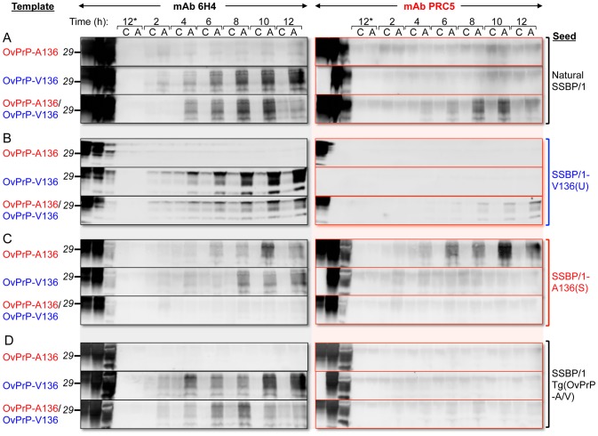
Figure 5. PMCA using defined seeds and substrates.
PMCA was performed for various times indicated. At each time point samples were either amplified by sonication (A), or matching control samples (C) received no sonication, and were therefore not amplified. Brain homogenates from healthy Tg(OvPrP-A136)3533+/− and Tg(OvPrP-V136)4166+/− mice served as sources of OvPrP-A136, OvPrP-V136 or mixtures of the two. In A, samples were seeded with sheep SSBP/1; in B, samples were seeded with brain extracts from diseased Tg(OvPrP-V136)4166+/− mice [SSBP/1-V136(U) prions]; in C, samples were seeded with brain extracts from diseased Tg(OvPrP-A136)3533+/− mice [SSBP/1-A136(S); in D, samples were seeded with extracts from diseased Tg(OvPrP-A/V) mice. Samples labeled 12* received no seed. The first three lanes of each immunoblot were loaded with the substrate(s) for each PMCA reaction, not treated with PK; the corresponding seed, not treated with PK; and, the same seed treated with PK. All other samples were digested with PK. Western blots were probed with mAbs 6H4 and PRC5 as indicated.

