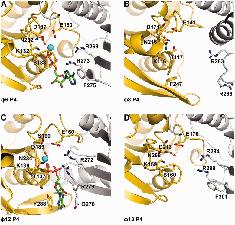
Figure 5.
Cartoon representation of the nucleotide binding sites of ɸ6 (A), ɸ8 (B), ɸ12 (C) and ɸ13 (D) P4s. Within hexamers, adjacent monomers are coloured in yellow and grey. Nucleotides (ADP), if present, are depicted as sticks with carbon atoms coloured in green. Oxygen, nitrogen and phosphorus atoms are coloured in red, blue and orange, respectively, and the position of Mg2+ (ɸ12 P4) or Ca2+ (ɸ6 P4) is indicated with a cyan sphere.

