Figure 5.
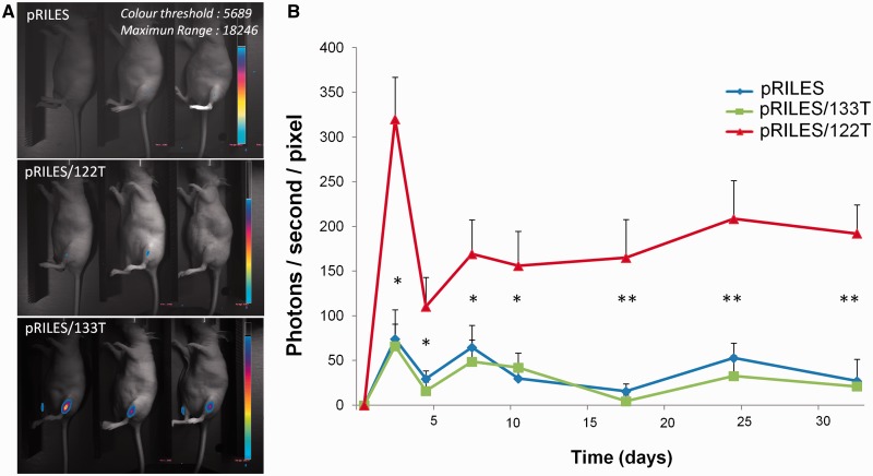
Kinetics of luciferase expression in the tibialis anterior muscle of immunodeficient mice. Two micrograms of pRILES/133T, pRILES/122T and control pRILES combined with 6 µg of the pQE30 empty expression plasmid were formulated with the 704 amphiphilic block copolymer and intramuscularly injected in the left and right tibialis anterior to transfect the skeletal muscles of nude mice. ROIs covering the lower legs of animals were drawn and light emission was quantified over time, from day 1 (before the intramuscular injection of RILES-derivates plasmids) to day 38, the end point of the assay. (A) Representative bioluminescence images collected at day 12 from three out of five mice per group. (B) Quantification of bioluminescence signals emitted from mice and plotted as a function of time. Error bars in B, mean ± SEM (n = 6) of one representative experiment repeated two times. Statistics by the two-tailed t-test, *P < 0.05; **P < 0.01 compared with the pRILES control group.

