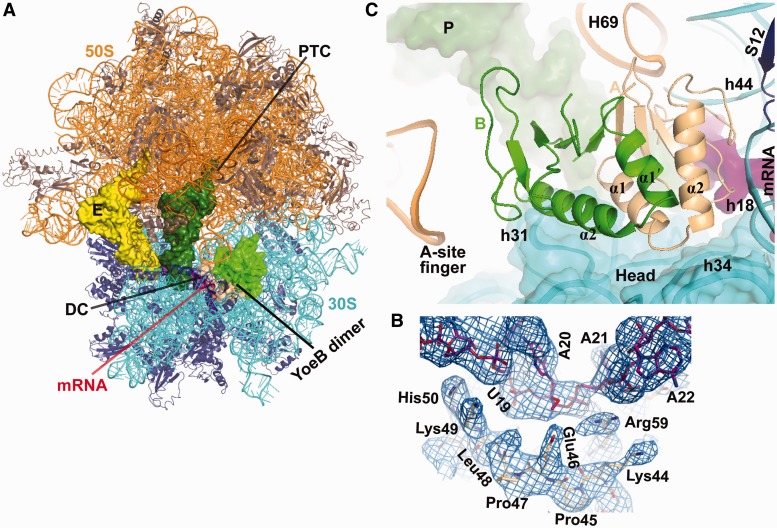
Figure 2.
Structure of YoeB bound to the 70S ribosome. (A) Overall view of the complex with YoeB dimer (colored lightorange for monomer A and green for monomer B). The peptidyl transferase center (PTC) and the decoding center (DC) are indicated. (B) Unbiased difference Fourier electron density map displayed at 1.2σwith refined YoeB and mRNA. (C) Close-up of YoeB binding site. The N-terminal two helices for both YoeB monomers A and B are labeled. The 16S head is shown as cartoon with transparent surface. RNA helices are numbered with E. coli sequence, prefixed by H for 23S rRNA and h for 16S rRNA.

