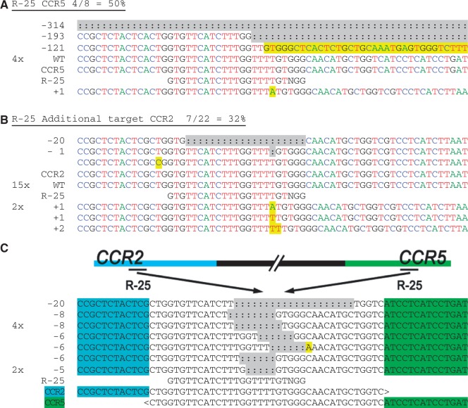
Figure 4.
Chromosomal deletions in CCR5 and CCR2 induced by CRISPR/Cas9 systems. HEK-293T cells were transfected with each CRISPR construct, and their genomic DNA harvested after 3 days in culture. The (a) on- and (b) off-target loci for guide strands R-25 were amplified with flanking PCR primers, cloned and Sanger sequenced. Sequencing reads are given for each guide strand and aligned to the wild-type sequence. The number of times each read occurred is indicated to the left of the alignment. Unmodified reads are indicated by ‘WT’. In (b) the guide strand mismatch is boxed. In (a) and (b), the A, C, T and G nucleotides are shown in green, blue, red and black, respectively, for clarity. (c) Genomic DNA from cells treated with R-25 was amplified using a CCR2 forward primer and reverse primer downstream of the CCR5 site. The PCR products were sequenced and aligned to ‘CCR2-CCR5’ with the bases unique to CCR2 or CCR5 indicated in blue or green, respectively, surrounding an identical area found in both genes. Sequencing detected that each product contained indels and mutations consistent with NHEJ, near the target sites for R-25. Insertions and point mutations are marked in yellow and deletions (:) are highlighted in gray.

