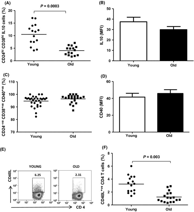
Fig. 2.

Impaired IL10 production by CD19+CD24hiCD38hi B cells on CD3 stimulation with age is due to impaired T-cell helper activity. PBMCs isolated from 15 healthy young and older adults were stimulated via CD3 for 72 h and stained for surface expression of CD19, CD24, CD38 and intracellularly for IL10. (A) Scatter plots show the percentage of IL10 positive cells within the CD19+CD24hiCD38hi B-cell subset in healthy young and old donors; (B) Bar charts showing the mean fluorescence intensity of IL10 staining within CD19+CD24hiCD38hi B cells after 72 h; (C) PBMCs isolated from 30 healthy young and older adults were stained for CD19, CD24, CD38 and CD40. Scatter plot shows the percentage of CD40+veCD24hiCD38hi B cells in unstimulated peripheral blood of healthy young and aged donors; (D). Bar chart comparing the expression of CD40 (MFI) on CD19+CD24hiCD38hi B cells without stimulation between young and old donors; (E) Representative flow cytometric plots showing the percentage of CD40L+ CD4T cells in healthy young and old donors after CD3 stimulation for 72 h; (F) Scatter plots showing the mean percentage of CD40L+ CD4T cells in PBMCs of 15 healthy young and older donors.
